Tag Archives: Health

DeepVariant: Highly Accurate Genomes With Deep Neural Networks


(Crossposted on the Google Open Source Blog)

Across many scientific disciplines, but in particular in the field of genomics, major breakthroughs have often resulted from new technologies. From Sanger sequencing, which made it possible to sequence the human genome, to the microarray technologies that enabled the first large-scale genome-wide experiments, new instruments and tools have allowed us to look ever more deeply into the genome and apply the results broadly to health, agriculture and ecology.

One of the most transformative new technologies in genomics was high-throughput sequencing (HTS), which first became commercially available in the early 2000s. HTS allowed scientists and clinicians to produce sequencing data quickly, cheaply, and at scale. However, the output of HTS instruments is not the genome sequence for the individual being analyzed — for humans this is 3 billion paired bases (guanine, cytosine, adenine and thymine) organized into 23 pairs of chromosomes. Instead, these instruments generate ~1 billion short sequences, known as reads. Each read represents just 100 of the 3 billion bases, and per-base error rates range from 0.1-10%. Processing the HTS output into a single, accurate and complete genome sequence is a major outstanding challenge. The importance of this problem, for biomedical applications in particular, has motivated efforts such as the Genome in a Bottle Consortium (GIAB), which produces high confidence human reference genomes that can be used for validation and benchmarking, as well as the precisionFDA community challenges, which are designed to foster innovation that will improve the quality and accuracy of HTS-based genomic tests.
For any given location in the genome, there are multiple reads among the ~1 billion that include a base at that position. Each read is aligned to a reference, and then each of the bases in the read is compared to the base of the reference at that location. When a read includes a base that differs from the reference, it may indicate a variant (a difference in the true sequence), or it may be an error.
Today, we announce the open source release of DeepVariant, a deep learning technology to reconstruct the true genome sequence from HTS sequencer data with significantly greater accuracy than previous classical methods. This work is the product of more than two years of research by the Google Brain team, in collaboration with Verily Life Sciences. DeepVariant transforms the task of variant calling, as this reconstruction problem is known in genomics, into an image classification problem well-suited to Google's existing technology and expertise.
Each of the four images above is a visualization of actual sequencer reads aligned to a reference genome. A key question is how to use the reads to determine whether there is a variant on both chromosomes, on just one chromosome, or on neither chromosome. There is more than one type of variant, with SNPs and insertions/deletions being the most common. A: a true SNP on one chromosome pair, B: a deletion on one chromosome, C: a deletion on both chromosomes, D: a false variant caused by errors. It's easy to see that these look quite distinct when visualized in this manner.
We started with GIAB reference genomes, for which there is high-quality ground truth (or the closest approximation currently possible). Using multiple replicates of these genomes, we produced tens of millions of training examples in the form of multi-channel tensors encoding the HTS instrument data, and then trained a TensorFlow-based image classification model to identify the true genome sequence from the experimental data produced by the instruments. Although the resulting deep learning model, DeepVariant, had no specialized knowledge about genomics or HTS, within a year it had won the the highest SNP accuracy award at the precisionFDA Truth Challenge, outperforming state-of-the-art methods. Since then, we've further reduced the error rate by more than 50%.
DeepVariant is being released as open source software to encourage collaboration and to accelerate the use of this technology to solve real world problems. To further this goal, we partnered with Google Cloud Platform (GCP) to deploy DeepVariant workflows on GCP, available today, in configurations optimized for low-cost and fast turnarounds using scalable GCP technologies like the Pipelines API. This paired set of releases provides a smooth ramp for users to explore and evaluate the capabilities of DeepVariant in their current compute environment while providing a scalable, cloud-based solution to satisfy the needs of even the largest genomics datasets.

DeepVariant is the first of what we hope will be many contributions that leverage Google's computing infrastructure and ML expertise to both better understand the genome and to provide deep learning-based genomics tools to the community. This is all part of a broader goal to apply Google technologies to healthcare and other scientific applications, and to make the results of these efforts broadly accessible.

Assisting Pathologists in Detecting Cancer with Deep Learning


A pathologist’s report after reviewing a patient’s biological tissue samples is often the gold standard in the diagnosis of many diseases. For cancer in particular, a pathologist’s diagnosis has a profound impact on a patient’s therapy. The reviewing of pathology slides is a very complex task, requiring years of training to gain the expertise and experience to do well.

Even with this extensive training, there can be substantial variability in the diagnoses given by different pathologists for the same patient, which can lead to misdiagnoses. For example, agreement in diagnosis for some forms of breast cancer can be as low as 48%, and similarly low for prostate cancer. The lack of agreement is not surprising given the massive amount of information that must be reviewed in order to make an accurate diagnosis. Pathologists are responsible for reviewing all the biological tissues visible on a slide. However, there can be many slides per patient, each of which is 10+ gigapixels when digitized at 40X magnification. Imagine having to go through a thousand 10 megapixel (MP) photos, and having to be responsible for every pixel. Needless to say, this is a lot of data to cover, and often time is limited.

To address these issues of limited time and diagnostic variability, we are investigating how deep learning can be applied to digital pathology, by creating an automated detection algorithm that can naturally complement pathologists’ workflow. We used images (graciously provided by the Radboud University Medical Center) which have also been used for the 2016 ISBI Camelyon Challenge1 to train algorithms that were optimized for localization of breast cancer that has spread (metastasized) to lymph nodes adjacent to the breast.

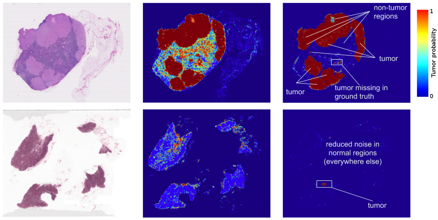
The results? Standard “off-the-shelf” deep learning approaches like Inception (aka GoogLeNet) worked reasonably well for both tasks, although the tumor probability prediction heatmaps produced were a bit noisy. After additional customization, including training networks to examine the image at different magnifications (much like what a pathologist does), we showed that it was possible to train a model that either matched or exceeded the performance of a pathologist who had unlimited time to examine the slides.
Left: Images from two lymph node biopsies. Middle: earlier results of our deep learning tumor detection. Right: our current results. Notice the visibly reduced noise (potential false positives) between the two versions.
In fact, the prediction heatmaps produced by the algorithm had improved so much that the localization score (FROC) for the algorithm reached 89%, which significantly exceeded the score of 73% for a pathologist with no time constraint2. We were not the only ones to see promising results, as other groups were getting scores as high as 81% with the same dataset. Even more exciting for us was that our model generalized very well, even to images that were acquired from a different hospital using different scanners. For full details, see our paper “Detecting Cancer Metastases on Gigapixel Pathology Images”.
A closeup of a lymph node biopsy. The tissue contains a breast cancer metastasis as well as macrophages, which look similar to tumor but are benign normal tissue. Our algorithm successfully identifies the tumor region (bright green) and is not confused by the macrophages.
While these results are promising, there are a few important caveats to consider.
  • Like most metrics, the FROC localization score is not perfect. Here, the FROC score is defined as the sensitivity (percentage of tumors detected) at a few pre-defined average false positives per slide. It is pretty rare for a pathologist to make a false positive call (mistaking normal cells as tumor). For example, the score of 73% mentioned above corresponds to a 73% sensitivity and zero false positives. By contrast, our algorithm’s sensitivity rises when more false positives are allowed. At 8 false positives per slide, our algorithms had a sensitivity of 92%.
  • These algorithms perform well for the tasks for which they are trained, but lack the breadth of knowledge and experience of human pathologists — for example, being able to detect other abnormalities that the model has not been explicitly trained to classify (e.g. inflammatory process, autoimmune disease, or other types of cancer).
  • To ensure the best clinical outcome for patients, these algorithms need to be incorporated in a way that complements the pathologist’s workflow. We envision that algorithm such as ours could improve the efficiency and consistency of pathologists. For example, pathologists could reduce their false negative rates (percentage of undetected tumors) by reviewing the top ranked predicted tumor regions including up to 8 false positive regions per slide. As another example, these algorithms could enable pathologists to easily and accurately measure tumor size, a factor that is associated with prognosis.
Training models is just the first of many steps in translating interesting research to a real product. From clinical validation to regulatory approval, much of the journey from “bench to bedside” still lies ahead — but we are off to a very promising start, and we hope by sharing our work, we will be able to accelerate progress in this space.


1 For those who might be interested, the Camelyon17 challenge, which builds upon the 2016 challenge, is currently underway.

2 The pathologist ended up spending 30 hours on this task on 130 slides.


Deep Learning for Detection of Diabetic Eye Disease


Diabetic retinopathy (DR) is the fastest growing cause of blindness, with nearly 415 million diabetic patients at risk worldwide. If caught early, the disease can be treated; if not, it can lead to irreversible blindness. Unfortunately, medical specialists capable of detecting the disease are not available in many parts of the world where diabetes is prevalent. We believe that Machine Learning can help doctors identify patients in need, particularly among underserved populations.

A few years ago, several of us began wondering if there was a way Google technologies could improve the DR screening process, specifically by taking advantage of recent advances in Machine Learning and Computer Vision. In "Development and Validation of a Deep Learning Algorithm for Detection of Diabetic Retinopathy in Retinal Fundus Photographs", published today in JAMA, we present a deep learning algorithm capable of interpreting signs of DR in retinal photographs, potentially helping doctors screen more patients in settings with limited resources.

One of the most common ways to detect diabetic eye disease is to have a specialist examine pictures of the back of the eye (Figure 1) and rate them for disease presence and severity. Severity is determined by the type of lesions present (e.g. microaneurysms, hemorrhages, hard exudates, etc), which are indicative of bleeding and fluid leakage in the eye. Interpreting these photographs requires specialized training, and in many regions of the world there aren’t enough qualified graders to screen everyone who is at risk.
Figure 1. Examples of retinal fundus photographs that are taken to screen for DR. The image on the left is of a healthy retina (A), whereas the image on the right is a retina with referable diabetic retinopathy (B) due a number of hemorrhages (red spots) present.
Working closely with doctors both in India and the US, we created a development dataset of 128,000 images which were each evaluated by 3-7 ophthalmologists from a panel of 54 ophthalmologists. This dataset was used to train a deep neural network to detect referable diabetic retinopathy. We then tested the algorithm’s performance on two separate clinical validation sets totalling ~12,000 images, with the majority decision of a panel 7 or 8 U.S. board-certified ophthalmologists serving as the reference standard. The ophthalmologists selected for the validation sets were the ones that showed high consistency from the original group of 54 doctors.

Performance of both the algorithm and the ophthalmologists on a 9,963-image validation set are shown in Figure 2.
Figure 2. Performance of the algorithm (black curve) and eight ophthalmologists (colored dots) for the presence of referable diabetic retinopathy (moderate or worse diabetic retinopathy or referable diabetic macular edema) on a validation set consisting of 9963 images. The black diamonds on the graph correspond to the sensitivity and specificity of the algorithm at the high sensitivity and high specificity operating points.
The results show that our algorithm’s performance is on-par with that of ophthalmologists. For example, on the validation set described in Figure 2, the algorithm has a F-score (combined sensitivity and specificity metric, with max=1) of 0.95, which is slightly better than the median F-score of the 8 ophthalmologists we consulted (measured at 0.91).

These are exciting results, but there is still a lot of work to do. First, while the conventional quality measures we used to assess our algorithm are encouraging, we are working with retinal specialists to define even more robust reference standards that can be used to quantify performance. Furthermore, interpretation of a 2D fundus photograph, which we demonstrate in this paper, is only one part in a multi-step process that leads to a diagnosis for diabetic eye disease. In some cases, doctors use a 3D imaging technology, Optical Coherence Tomography (OCT), to examine various layers of a retina in detail. Applying machine learning to this 3D imaging modality is already underway, led by our colleagues at DeepMind. In the future, these two complementary methods might be used together to assist doctors in the diagnosis of a wide spectrum of eye diseases.

Automated DR screening methods with high accuracy have the strong potential to assist doctors in evaluating more patients and quickly routing those who need help to a specialist. We are working with doctors and researchers to study the entire process of screening in settings around the world, in the hopes that we can integrate our methods into clinical workflow in a manner that is maximally beneficial. Finally, we are working with the FDA and other regulatory agencies to further evaluate these technologies in clinical studies.

Given the many recent advances in deep learning, we hope our study will be just one of many compelling examples to come demonstrating the ability of machine learning to help solve important problems in medical imaging in healthcare more broadly.

Learn more about the Health Research efforts of the Brain team at Google

Reproducible Science: Cancer Researchers Embrace Containers in the Cloud


Today we hear from the principal investigators of the ICGC-TCGA DREAM Somatic Mutation Calling Challenges about how they are encouraging cancer researchers to make use of Docker and Google Cloud Platform to gain a deeper understanding of the complex genetic mutations that occur in cancer, while doing so in a reproducible way.
– Nicole Deflaux and Jonathan Bingham, Google Genomics

Today’s genomic analysis software tools often give different answers when run in different computing environments - that’s like getting a different diagnosis from your doctor depending on which examination room you’re sitting in. Reproducible science matters, especially in cancer research where so many lives are at stake. The Cancer Moonshot has called for the research world to 'Break down silos and bring all the cancer fighters together'. Portable software “containers” and cloud computing hold the potential to help achieve these goals by making scientific data analysis more reproducible, reusable and scalable.

Our team of researchers from the Ontario Institute for Cancer Research, University of California Santa Cruz, Sage Bionetworks and Oregon Health and Sciences University is pushing the frontiers by encouraging scientists to package up their software in reusable Docker containers and make use of cloud-resident data from the Cancer Cloud Pilots funded by the National Cancer Institute.

In 2014 we initiated the ICGC-TCGA DREAM Somatic Mutation Calling (SMC) Challenges where Google provided credits on Google Cloud Platform. The first result of this collaboration was the DREAM-SMC DNA challenge, a public challenge that engaged cancer researchers from around the world to find the best methods for discovering DNA somatic mutations. By the end of the challenge, over 400 registered participants competed by submitting 3,500 open-source entries for 14 test genomes, providing key insights on the strengths and limitations of the current mutation detection methods.

The SMC-DNA challenge enabled comparison of results, but it did little to facilitate the exchange of cross-platform software tools. Accessing extremely large genome sequence input files and shepherding complex software pipelines created a “double whammy” to discourage data sharing and software reuse.

How can we overcome these barriers?

Exciting developments have taken place in the past couple of years that may annihilate these last barriers. The availability of cloud technologies and containerization can serve as the vanguards of reproducibility and interoperability.

Thus, a new way of creating open DREAM challenges has emerged: rather than encouraging the status quo where participants run their own methods themselves on their own systems, and the results cannot be verified, the new challenge design requires participants to submit open-source code packaged in Docker containers so that anyone can run their methods and verify the results. Real-time leaderboards show which entries are winning and top performers have a chance to claim a prize.

Working with Google Genomics and Google Cloud Platform, the DREAM-SMC organizers are now using cloud and containerization technologies to enable portability and reproducibility as a core part of the DREAM challenges. The latest SMC installments, the SMC-Het Challenge and the SMC-RNA Challenge have implemented this new plan:

  • SMC-Het Challenge: Tumour biopsies are composed of many different cell types in addition to tumour cells, including normal tissue and infiltrating immune cells. Furthermore, the tumours themselves are made of a mixture of different subpopulations, all related to one another through cell division and mutation. Critically, each sub-population can have distinct clinical outcomes, with some more resistant to treatment or more likely to metastasize than others. The goal of the SMC-Het Challenge is to identify the best methods for predicting tumor subpopulations and their “family tree” of relatedness from genome sequencing data.
  • SMC-RNA Challenge: The alteration of RNA production is a fundamental mechanism by which cancer cells rewire cellular circuitry. Genomic rearrangements in cancer cells can produce fused protein products that can bestow Frankenstein-like properties. Both RNA abundances and novel fusions can serve as the basis for clinically-important prognostic biomarkers. The SMC-RNA Challenge will identify the best methods to detect such rogue expressed RNAs in cancer cells.

Ultimately, the success will be gauged by the amount of serious participation in these latest competitions. So far, the signs are encouraging. SMC-Het, which focuses on a very new research area, launched in November 2015 and has already enlisted 18 teams contributing over 70 submissions. SMC-RNA just recently launched and will run until early 2017, with several of the world leaders in the field starting to prepare entries. What’s great about the submissions being packaged in containers is that even after the challenges end, the tested methods can be applied and further adapted by anyone around the world.

Thus, the moon shot need not be a lucky solo attempt made by one hero in one moment of inspiration. Instead, the new informatics of clouds and containers will enable us to combine intelligence so we can build a series of bridges from here to there.

To participate in the DREAM challenges, visit the SMC-Het and SMC-RNA Challenge sites.